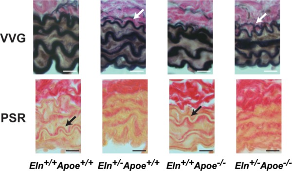
Fig. 5.

Representative carotid artery sections stained with VVG (top) and PSR (bottom) for groups on WD. VVG stains elastic fibers black, muscle brown, and collagen pink. PSR stains collagen red on a pale yellow background. (Please refer to online article for color figures.) Note the additional layer of elastic fibers in Eln+/− VVG images (white arrows) and the clear outline of collagen fibers around the elastic fibers in Eln+/+ PSR images (black arrows). Scale bars = 10 μm.
