Figure 1.

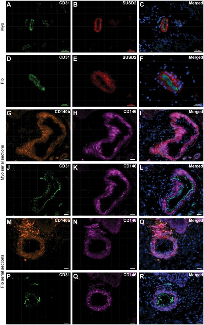
Localisation of SUSD2 + and CD146 + CD140b + cells in myometrium and fibroid tissues. Immunofluorescence analyses were performed on frozen tissue sections to localise putative MSC markers. SUSD2 expression (B, E) is primarily localised to the perivascular region surrounding CD31+ endothelial cells (A, D) in myometrial (A–C) and fibroid (D–F) tissue. (G–I) CD146 and CD140b co-expressing cells in myometrium tissue. (J–L) serial section to (G–I) showing CD31+ endothelial cells with surrounding CD146+ perivascular cells. (M–O) CD146 and CD140b co-expressing cells in fibroid tissue and (P–R) serial section showing CD31+ endothelial cells with surrounding CD146+ perivascular cells. Scale bars are 25 μM; nuclei are shown stained with DAPI in merged images, which are representative of n = 5 patient samples.
