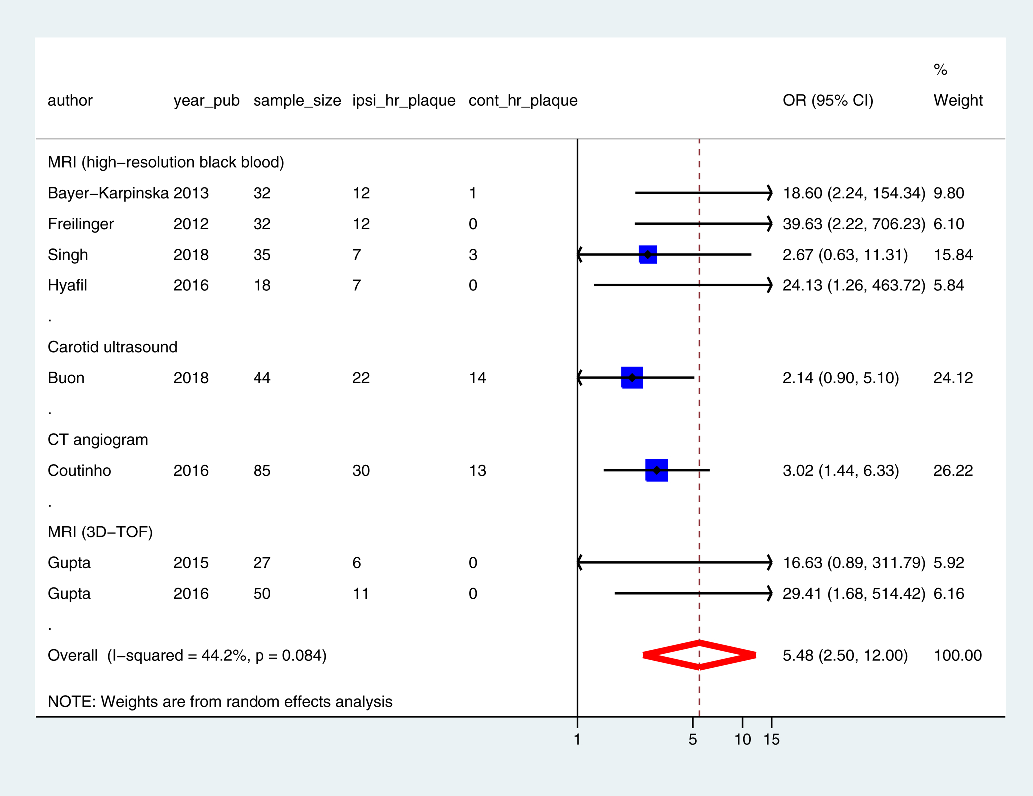
Figure 2: Odds-ratio of finding plaque with high-risk features in the ipsilateral versus the contralateral carotid in ESUS.

3D-TOF = 3-dimensional time of flight, CI = Confidence interval, CT = Computed tomography, cont_hr_plaque = contralateral carotid plaque with high-risk features, ipsi_hr_plaque = ipsilateral carotid plaque with high-risk features, MRI = Magnetic resonance imaging, OR = Odds ratio, sample_size = number of participants in the study, year_pub = year of publication
