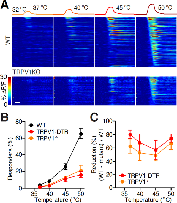
Figure 3 |. Role of TRPV1 channel and TRPV1-expressing DRG neurons in sensing warmth.
A. Heat maps of the activities of all heat-responding neurons in representative fields-of-view (FOVs) in an example wild type (WT, 396 neurons) and TRPV1−/− (227 neurons) mouse. Neurons were considered responders when the maximum ΔF/F of each individual trial exceeded 5% and 2.5 times of σ0 (s.d. of the prestimulus baseline) above F0 of each individual trial, and the maximum ΔF/F of the averaged and smoothed response exceeded 5% and 3 times σ0 above F0 of the response averaged from all the trials of the same stimulus. From detailed methods, see Ran et al., 2016. Scale bar, 10 s.
B. Temperature-response relationship in WT and TRPV1−/− mice. (Black: WT, n = 10 mice; orange: TRPV1−/−, n = 4 mice; red: TRPV1-DTR, n = 9 mice. WT and TRPV1-DTR data are from Ran et al., 2016. Mann-Whitney test between TRPV1−/− and WT, P = 0.053, P = 0.024, P = 0.008, P = 0.004 for the four temperatures, respectively.)
C. The percentage of reduction (calculate from B) of heat-responding neurons in TRPV1−/− mice compared to WT.

