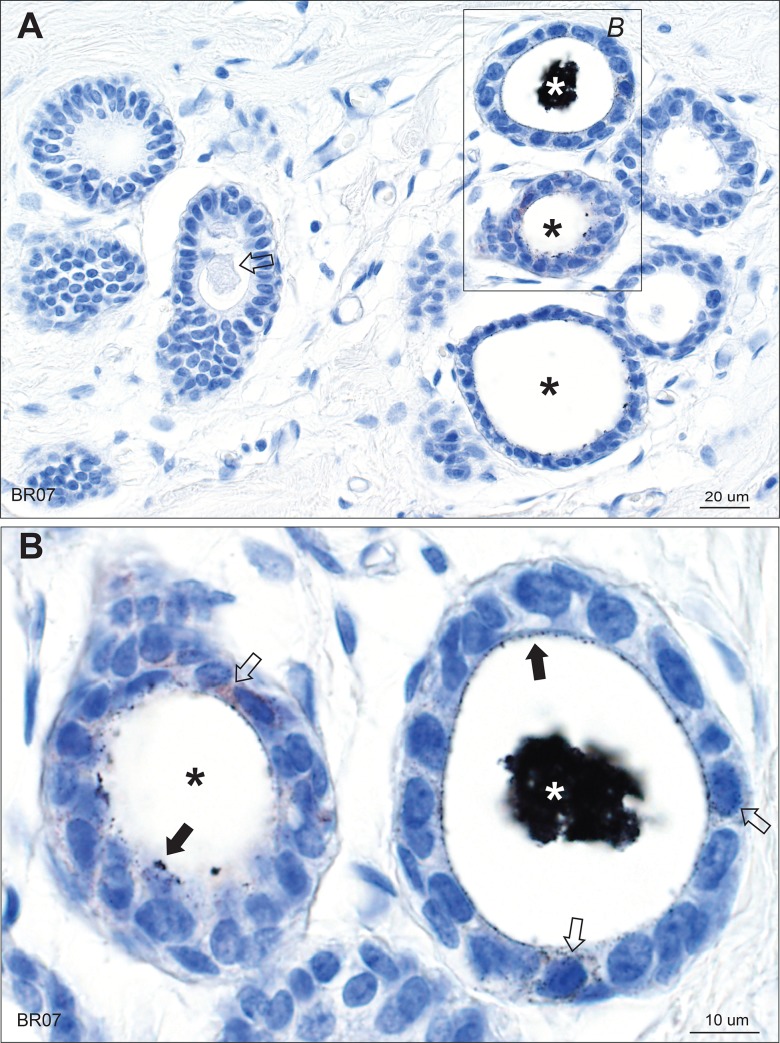
Fig 1. Mercury in breast lobules.
(A) One lobule in the group on the right shows black mercury-stained secretion (white asterisk), and two (black asterisks) show mercury on the luminal surface of epithelial cells. The lobules on the left show no mercury, with unstained luminal secretion visible in one (arrow). (B) Enlarged image of the box in A. The lobule on the right has mercury in its artefactually-shrunken luminal secretion (white asterisk). Some mercury-stained secretion is still attached to the luminal surface of the epithelial cells (solid arrows). Fine particulate mercury staining is present in some epithelial cells (open arrows). In the lobule on the left (black asterisk) much of the secretion has fallen out during processing, but mercury-stained secretion remains attached to the luminal surface of some epithelial cells (closed arrow) and is also present in some epithelial cells (open arrow). AMG/hematoxylin. BR: sample identity number.

