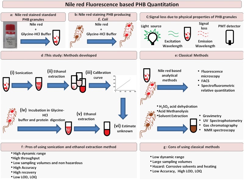
Figure 1:
Schematic workflow. Pictorial representation of workflows/methods for improved accuracy of quantitation of PHB. The principle of the assay is depicted in Panels a–c. Standard PHB suspensions in aqueous glycine-HCl buffer bind quantitatively to Nile red and have specific fluorescence spectral characteristics. Panel b shows how cells producing PHA can be, grown in the presence of Nile red that can bind to intracellular carbonosome, an intracellular PHA producing inclusion body. Panel c shows loss in emission signal of fluorescence dependent on physical properties of granules. Panel d represents the methodology developed in this study while panel e represents the classical method for quantitation using UV spectrophotometry. Panels f–g discuss the Pros and Cons of the methods.

