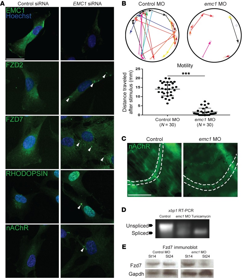
Figure 4. Knockdown of EMC1 in human RPE cells and Xenopus affects multiple transmembrane proteins and embryo motility and activates ER stress.
(A) Immunofluorescence antibody labeling of EMC1 revealed a decrease in its expression after EMC1 siRNA treatment as compared with control siRNA treatments. Multipass membrane proteins (RHODOPSIN, nAChR, FZD2, FZD7) were abnormally localized (n = 10 high power fields per marker per condition done in 3 replicates). Scale bar: 20 μm. (B) Sample traces and measurement of control morphant (n = 30) and emc1 morphant (n = 30) tadpole movement over 10 seconds after stimulation (different colors differentiate distinct tadpoles) over 3 replicates. (C) Labeling of nAChR in the proximal tail of emc1-depleted stage 45 tadpoles showed sparse and less intense expression as compared with control counterparts. Scale bar: 50 μm. (D) Splicing assay for xbp1 in pooled (n = 30 per condition) stage 24 Xenopus embryos displayed increased splicing with emc1 MO depletion compared with control embryos repeated in 4 biological replicates. Tunicamycin treatment acted as positive control. (E) Immunoblotting for Fzd7 showed similar levels of Fzd7 in pooled (n = 30 per stage per condition) emc1 morphants as compared with control morphants at stage 14, but a marked decrease in levels at stage 24. ***P < 0.0005 by Student’s t test. Bars indicate mean and SD.

