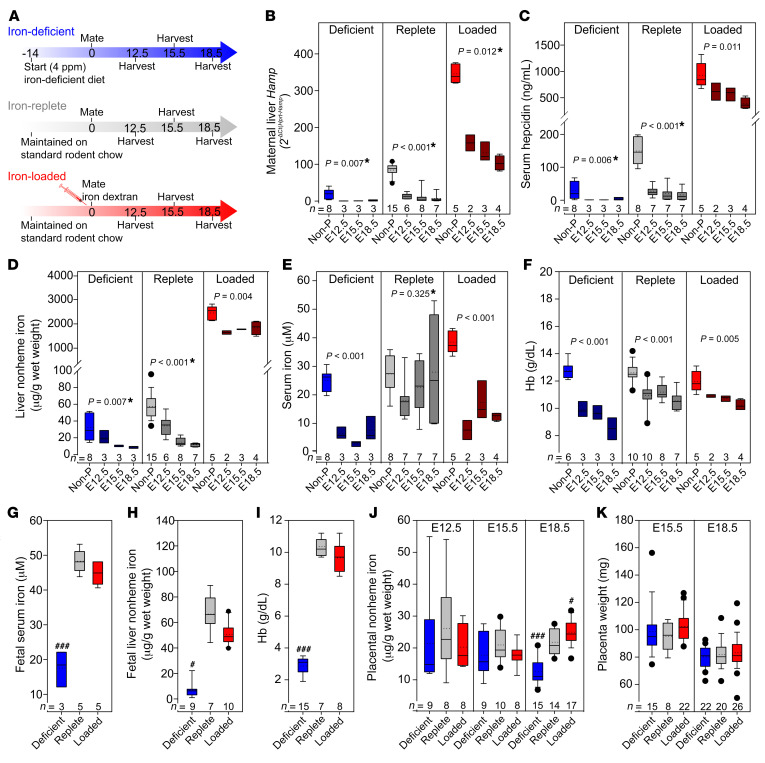
Figure 1. Maternal hepcidin and serum iron levels determine embryo and placental iron status.
The iron status of WT C57BL/6 female mice was altered using diet or iron dextran injections. (A) Adult females were fed standard chow (185 ppm iron) or a low-iron diet (4 ppm iron) ad libitum 2 weeks prior to and throughout pregnancy, or were injected with 20 mg iron dextran at the time of mating. Pregnant females were analyzed at E12.5, E15.5, and E18.5. Nonpregnant (Non-P) females were subjected to an equivalent iron treatment. (B–F) Maternal measurements of (B) hepcidin (Hamp) mRNA and (C) serum hepcidin. (D) Liver nonheme iron. (E) Serum iron concentration. (F) Hb concentration. Statistical differences between groups was determined by 1-way ANOVA for normally distributed values, or otherwise by 1-way ANOVA on ranks (indicated by a single asterisk after the P value). (G–I) Embryo measurements at E18.5 for (G) serum iron and (H) liver nonheme iron. (I) Hb concentration. (J) Placental nonheme iron levels at E12.5, E15.5, and E18.5. (K) Placental weight at E15.5 and E18.5 (we did not obtain whole placentas at E12.5). Statistical differences between groups was determined by 1-way ANOVA for normally distributed values followed by the Holm-Sidak method for multiple comparisons versus the iron-replete control group (###P < 0.001) or 1-way ANOVA on ranks followed by Dunn’s method for multiple comparisons versus the iron-replete control group (#P < 0.05). The numbers of animals are indicated in the x axes of the box and whisker plots.

