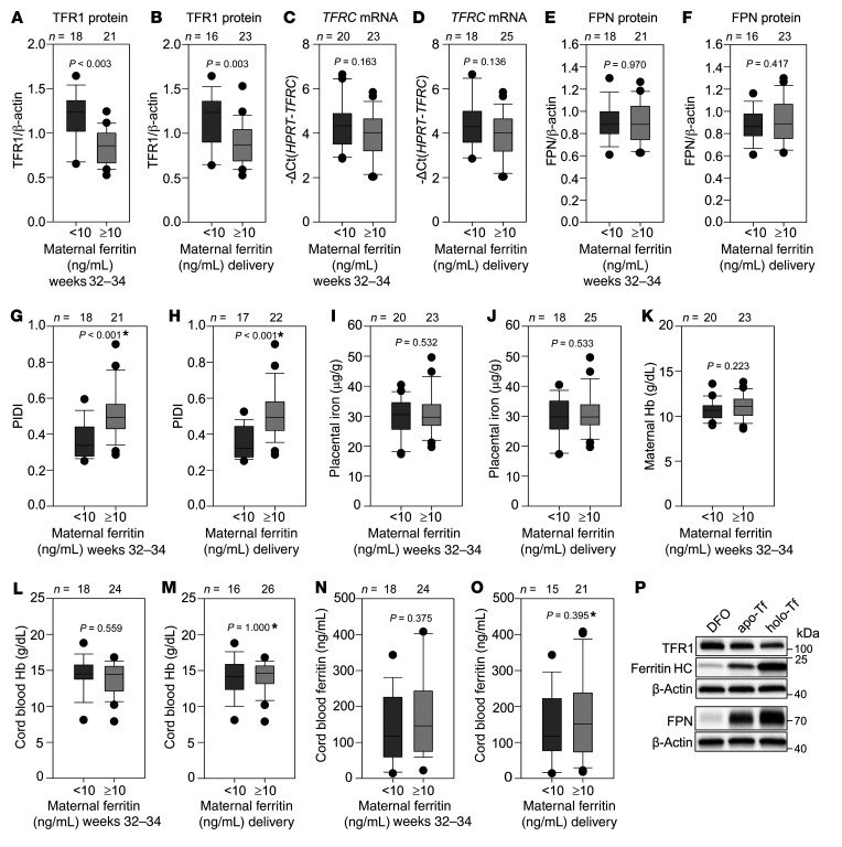
Figure 4. Placental response to maternal iron deficiency in human pregnancy.
Placentas from uncomplicated human pregnancies were analyzed by Western blotting to determine protein expression of TFR1 and FPN, normalized to β-actin. qPCR was performed to determine TFRC mRNA expression, normalized to HPRT. (A and B) TFR1 protein levels, (C and D) TFRC mRNA levels, and (E and F) FPN protein levels according to maternal ferritin during weeks 32–34 or at delivery. (G and H) The PIDI was calculated as the ratio of expression of placental FPN to TFR1 protein, with a lower PIDI reflecting pregnancies at increased risk of fetal iron deficiency. The PIDI was lower in pregnant women with serum ferritin levels below 10 ng/mL than in those with ferritin levels above 10 ng/mL, regardless of whether ferritin was measured at 32–34 weeks of pregnancy or at delivery. No difference between <10 ng/mL and >10 ng/mL ferritin groups was observed for (I and J) placental nonheme iron concentrations, (K) maternal Hb, (L and M) cord blood Hb, or (N and O) cord blood ferritin. (P) PHTs were treated with 100 μM DFO, apo-Tf or holo-Tf for 24 hours. TFR1, FPN, ferritin heavy chain (HC), and β-actin expression was assessed by Western blotting. Statistical differences between groups was determined by 2-tailed Student’s t test or Mann-Whitney U rank-sum test for non-normally distributed values (denoted by an asterisk after the P value). The numbers of animals are indicated above the box and whisker plots.

