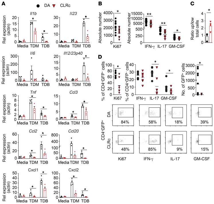
Figure 4. Attenuated response to Mcl/Mincle stimulation in CLRc BMMas resulting in altered CD4+ T cell extravasation and activation.
(A) Bone marrow–derived macrophages (BMMas) from DA (n = 4) and CLRc (n = 4) rats were stimulated with receptor-specific ligands (TDM and TDB) for 18 hours (representative of 3 experiments). Genes downstream of the Mcl/Mincle pathway were analyzed by qPCR. (B) MOG-specific CD4+ effector cells were reactivated for 4 days with DA (n = 6) or CLRc (n = 6) BMMas at a ratio of 40:1 (T cell/BMMa) in the presence of MOG peptide. Flow cytometry analysis of CD4+ T cell assessing cytokine production and proliferation (representative of 2 experiments). (C) Transendothelial extravasation of MOG-specific CD4+ effector cells toward DA (n = 4) or CLRc (n = 4) BMMas. Flow cytometry analysis of CD4+ T cell assessing transmigration (representative of 2 experiments). (D) Adoptive transfer of GFP+ MOG-specific CD4+ effector cells injected i.v. into DA (n = 7) or CLRc (n = 7) recipients 6 days p.i. (representative of 2 experiments). Characterization of proliferation and cytokine production in both GFP– as well as GFP+ infiltrating cells isolated from spinal cord on day 13 p.i. stimulated in vitro with PMA/ionomycin/brefeldin A for 5 hours. Data are presented as the mean ± SEM. All comparisons were analyzed with the Mann-Whitney U test. *P < 0.05; **P < 0.01.

