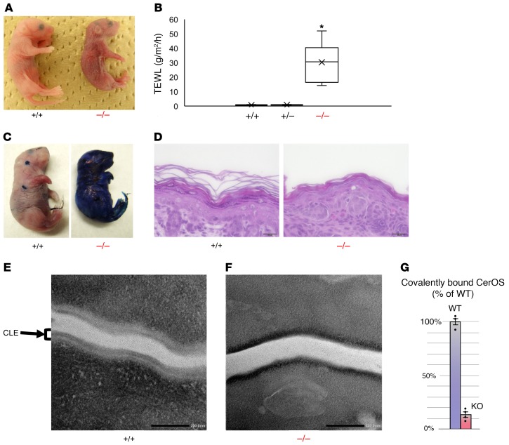
Figure 2. Sdr9c7 deletion reduces permeability barrier function and CLE formation.
(A) Gross appearance of Sdr9c7+/+ and Sdr9c7–/– newborns at 1 hour after birth. Although the newborns (Sdr9c7+/+, Sdr9c7+/–, Sdr9c7–/–) were indistinguishable at birth, Sdr9c7–/– mice show erythema and wrinkled skin consistent with increased TEWL soon after birth. (B) Skin permeability as assessed by TEWL on the dorsal skin surface of Sdr9c7+/+ (n = 6), Sdr9c7+/– (n = 7), and Sdr9c7–/– (n = 10) mice (mean ± SEM, *P < 0.001). (C) Toluidine blue exclusion assay using neonatal Sdr9c7+/+ and Sdr9c7–/– mice. (D) Histology of dorsal skin sections from newborn Sdr9c7+/+ and Sdr9c7–/– mice stained with hematoxylin and eosin. Scale bars: 20 µm. (E and F) Transmission electron microscopy of the stratum corneum in Sdr9c7+/+ and Sdr9c7–/– newborn mice. Scale bars = 200 μm. (E) There is a normal external lipid membrane monolayer, the corneocyte-bound lipid envelope (CLE), in Sdr9c7+/+ mice. (F) In contrast, the CLE is either absent, partially formed irregularly, or loosely attached to the CCE in Sdr9c7–/– mice. (G) The levels of covalently bound CerOS are significantly reduced in the epidermis of Sdr9c7–/– (KO) mice compared with that of Sdr9c7+/+ (WT) mice (mean ± SEM, n = 3 or 4, Student’s t test, P < 0.001).

