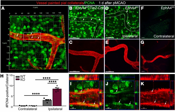
Figure 3. Increased cell proliferation within the MCA-ACA collateral niche of KO mice 1 day after pMCAO.
Vessel painted brains were immuno-labeled with anti-PCNA (green; white arrows) and imaged by high magnification confocal imaging. Maximum Z-projection analysis of the entire Z-stacked vessel was used to assess cell division for evidence of collateral remodeling as early as 1 day after pMCAO in the ipsilateral (A–E) and contralateral (F and G) hemispheres. (A) Representative 3D projected image from vessel painted (red) ipsilateral KO pial surface of an MCA-ACA collateral colabeled with antibodies against PCNA. KO collateral vessels show a greater number of PCNA+ cells aligned within (elongated compared with surrounding PCNA+ cells) the vessel wall compared with WT. (B and C) Representative single channel images of A, showing PCNA expression in the KO collateral niche compared with the ipsilateral WT collateral niche (D and E). No PCNA staining was seen in the contralateral pial collaterals of the same representative injured WT animal (F and G). (H) Quantified data showing the number of PCNA+ cells within the collateral vessel wall in increased in KO mice. (I–K) High-magnification confocal ortho view from inset in A, showing cell division in the KO collateral wall. Scale bars: 50 μm (B–G) and 10 μm (I–K). Two-way ANOVA with Bonferroni’s post hoc test; n = 5 mice per group; 4–5 MCA-ACA collaterals were analyzed per mouse. ****P < 0.0001.

