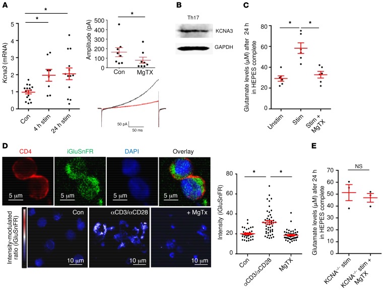
Figure 5. Th17 cells produce glutamate in a KV1.3-mediated manner.
(A) Kcna3 mRNA expression was upregulated after 4 hours and 24 hours of TCR stimulation with anti-CD3 and anti-CD28 (n = 17). Patch-clamp experiments were performed on Th17 cells and showed blockade of KV1.3 currents after application of MgTX (n = 9). (B) Western blot staining for KCNA3 and GAPDH was performed in Th17 cells (1 representative example of 4 is shown). (C) Glutamate levels after 24 hours in culture media were assessed, comparing TCR-stimulated Th17 cells with or without MgTX treatment (n = 6 per group). (D) Th17-differentiated cells were transfected with the GFP-based glutamate sensor iGluSnFR. Staining for CD4 was also performed, and GFP intensity was analyzed with ImageJ software. Scale bars: 5 μm (top row) and 10 μm (bottom row). Data indicate the mean ± SEM. (E) Glutamate levels were not significantly different between stimulated Th17 cells from WT and KCNA3–/– mice treated with MgTX (n = 3–4 per group). Data indicate the mean ± SEM. *P < 0.05, by 1-way ANOVA with Dunnett’s post hoc test (A, C, and D) or Mann-Whitney U test (E).

