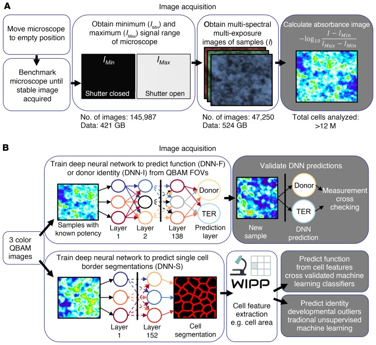
Figure 1. Methodology for image acquisition and image analysis.
(A) An overview of QBAM imaging. The method converts pixel values in bright-field images to absorbance values. (B) Methods of analyzing QBAM images of human iPSC-RPE to predict function, identity and developmental outliers. Three DNNs were constructed for this study: (a) Predicts function (TER and VEGF-ratio) from QBAM images (DNN-F) using entire FOVs from microscopes, (b) identifies whether QBAM images from different clones came from the same donor (DNN-I), (c) segments individual iPSC-RPE cells in absorbance images (DNN-S). QBAM images of iPSC-RPE that were segmented with DNN-S also had individual cell image features extracted with the WIPP, and these features were used to predict cell function, cell identity, and whether cells were developmental outliers. More information on QBAM imaging is presented in Supplemental Figure 1, and the DNN architectures are presented in Supplemental Figure 2. Features extracted by WIPP are listed in Supplemental Table 1.

