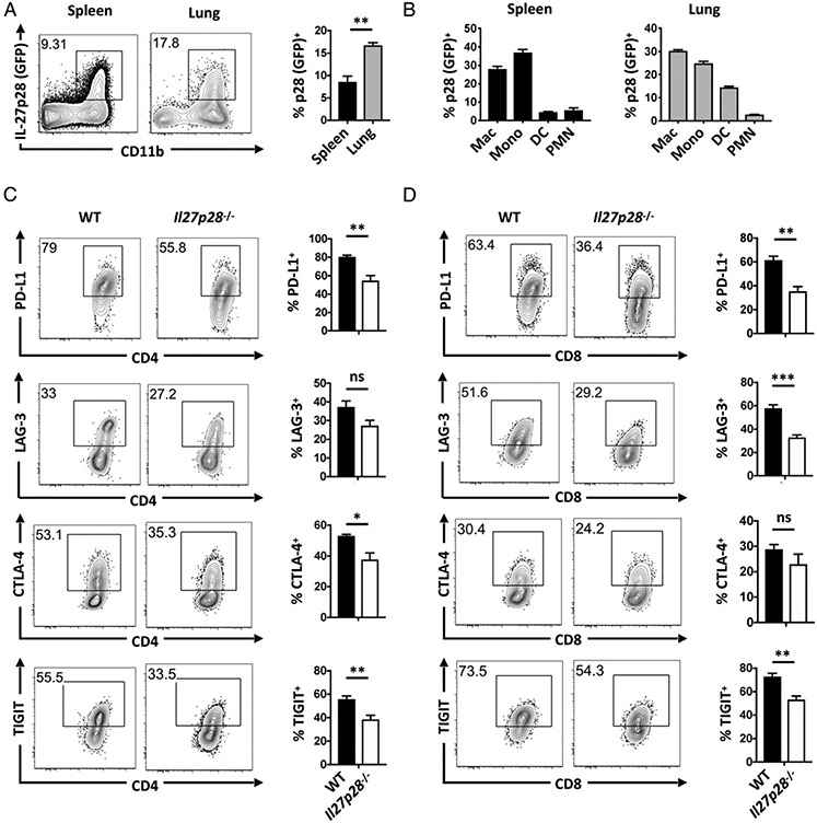
FIGURE 7. IL-27 contributes to IR expression by lung T cells during toxoplasmosis.
(A) Il27p28-GFP reporter mice were infected with 20 Me49 cysts i.p. for 10 d. p28-GFP was examined on nonlymphocytes (CD3−NK1.1−B220− CD19−) within the spleen (black bars) and lung (gray bars). (B) Within nonlymphocytes, p28-GFP expression was examined in macrophages (CD11b+CD64+Ly6GloF4/80+Ly6Clo), monocytes (CD11b+CD64+Ly6GloLy6Chi), DC (CD64−CD11c+MHCII+), and polymorphonuclear cells (CD11b+CD11c−CD64−Ly6cintLy6G+) in the spleen (left) and lung (right). (C and D) WT and Il27p28−/− mice were infected with 20 Me49 cysts i.p. for 11–12 d. IR expression by WT (black bars) and Il27p28−/− (white bars) lung tetramer+ CD4+ (B) and CD8+ (C) T cells. Three to five mice per group. Data representative of one (A) or four (B and C) independent experiments. Error bars indicate SEM. *p < 0.05, **p < 0.01, ***p < 0.001.

