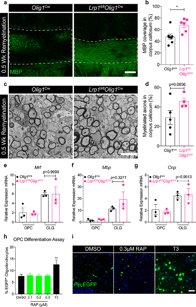
Fig. 4. Enhanced remyelination in mice lacking oligodendroglial LRP1 is not cell-autonomous.
(a) Representative images and (b) quantification of MBP expression in the remyelinating corpus callosum of Olig1cre and Lrp1fl/flOlig1cre mice after 0.5 Wk Rem (unpaired t-test; 2 independent experiments combined; scale bar 100μm). (c) Representative TEM images and (d) quantification of axons in the remyelinating corpus callosum after 0.5 Wk Rem (unpaired t-test; n=4–5 mice per genotype; scale bar 2μm). (e-g) qPCR for Mrf, Mbp, and Cnp from primary OPCs cultured in proliferation (OPC) or differentiation (OLG) media (unpaired t-test; n=3 mice per genotype). (h) Quantification and (i) representative images of Plp-EGFP OPCs treated with DMSO, T3, or increasing concentrations of the specific LRP1 inhibitor, RAP (One-way ANOVA with Dunnett’s multiple comparisons test, DMSO vs T3 **p=0.0072; conditions plated in sextuplicate; error bars represent +/− SEM; scale bar 150μm).

