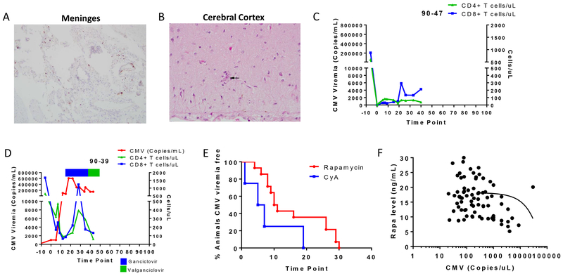
Figure 2. Effects of CMV viremia and reactivation timing.
Histological findings in the (A) meninges and (B) cerebral cortex of a CMV+ recipient (90-47) that developed uncontrollable CMV disease. C) CD4+ T cell (green) and CD8+ T cell (blue) counts for 90-47. D) CMV level (red), CD4+ T cell (green), and CD8+ T cell (blue) counts of recipient 90-39. Antiviral treatment with ganciclovir (blue bar) and valganciclovir (green bar) is indicated at the top of the graph. E) CMV reactivation time point for recipients receiving rapamycin (red, n=14) or CyA (blue, n=5) as immunosuppressant. F) Correlation between the rapamycin level and the day of CMV reactivation posttransplant.

