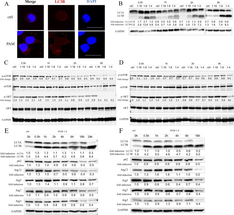
Figure 3.
Plasma-activated medium (PAM) induces autophagy of endometrial cancer cells. (A) Representative images of Immunocytochemistry staining by LC3B with or without PAM treatment for AMEC cells. (B) Expression levels of LC3 protein after 0.5–6 h PAM treatment in AMEC cells were evaluated by Western blotting in different concentrations. (C,D) Effect of PAM on mTOR and AKT activation after 0.5–6 h in different PAM concentrations in AMEC (C) and HEC50 (D) cells. (E,F) Expression levels of LC3, p62 and ATG family proteins after 1:4 PAM treatment in AMEC (E) and HEC50 (F) cells at 0–24 h. The numbers indicated the density value normalized to GAPDH using the ImageJ software. The density value of phosphorylated form was expressed as the ratio to the density value of total protein. The original blots are presented in Supplementary Fig. 3.

