Fig. 5.
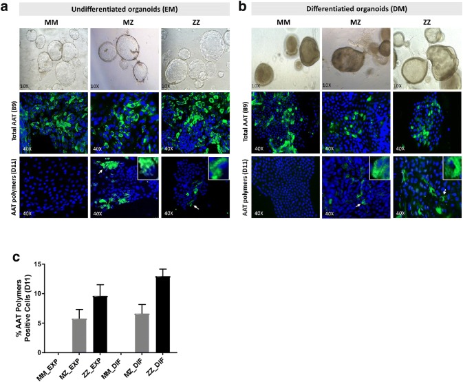
Immunofluorescence detection of AAT and AAT polymers. a Representative pictures of liver organoids in the expansion medium (EM) of MM, MZ and ZZ patients (images magnification ×10). b Differentiated liver organoids from MM, MZ and ZZ patients. Specific detection of total AAT protein with anti-AAT-B9 or with anti-AAT-D11 against AAT polymers, are shown in green fluorescence. Zoomed images of individual positive cells are showed in the right corner. c Quantification of AAT polymers (D11) positive cells in the different organoids MM, MZ and ZZ. Microscope Zeiss Ax10, camera Axio Cam Mrm Carl Zeiss and software AxioVision Rel.4.7 (images magnification ×40)

