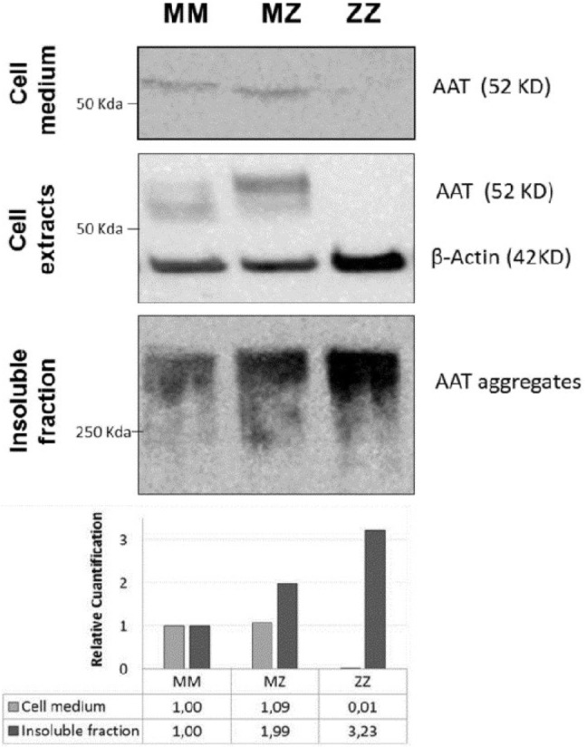
Fig. 6.

Representative western blot detection of AAT protein expression and secretion in MM, MZ and ZZ differentiated organoids. Blots of extracellular, cytoplasmic and insoluble fractions were incubated with anti-AAT-B9. The anti-β actin antibody was used as a control marker in the cytoplasm extract. Panels of figures were composed to show MM, MZ and ZZ AAT in the same order. Relative quantification with respect to MM amount of AAT in the extracellular medium and in the insoluble fraction of organoids is shown at the bottom
