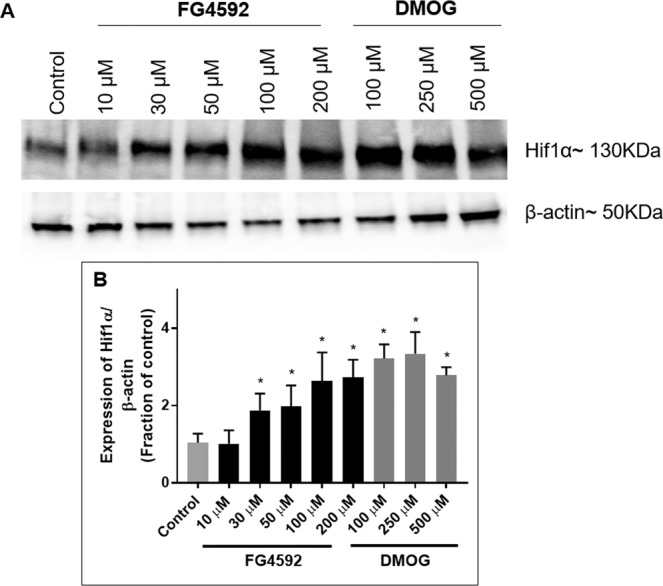
Figure 11.

Effects of HIF-PHD inhibitors on HIF-1α levels in primary rat neurons. Immunoblot analysis of HIF-1α levels in primary cortical neurons treated with 24 hours with 1% DMSO, FG4592 (10, 30 50, 100 µM) and DMOG (100, 250 µM) in normoxia. (A) Representative HIF-1α immunoblots were shown with those for β-actin; (B) Graph (n = 3) showed the normalised HIF-1α level measured at 24 hours after exposure to FG4592 or DMOG in normoxia. FG4592 (30 µM onwards) and DMOG (100, 250 µM) significantly increased HIF-1α levels in primary rat neurons. Error bars represent ± S.D. *Indicates P < 0.05 against control (1% DMSO treatment) (Two-way ANOVA, Tukey’s post-hoc analysis).
