Figure 4.
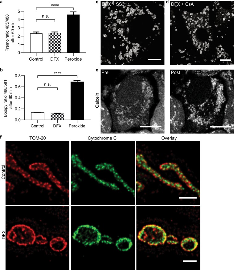
Deferasirox-induced swelling is independent of the mitochondrial permeability transition pore. (a) Cellular levels of ROS were measured using the ratiometric sensor Orp1-roGFP after 1 hour of treatment, and were not elevated in DFX (200 µM) treated OK cells. H2O2 was added directly to cells as a positive control (n = 4 experiments, total of 32 cells, mean ± SEM, one-way ANOVA with Tukey’s multiple comparisons test, ****p < 0.0001). (b) Lipid peroxidation after 1 hour treatment was measured using Bodipy 581/591 C11, and was also not increased in DFX (200 µM) treated cells (n = 4 experiments, total of 16 cells, mean ± SEM, one-way ANOVA with Tukey’s multiple comparisons test, ****p < 0.0001). (c,d) Neither the antioxidant SS-31 (500 µM) nor the mPTP inhibitor cyclosporin A (CsA, 1 µM) prevented subsequent DFX-induced mitochondrial swelling (scale = 10 µm). (e) Calcein signal remained localized to mitochondria after 25 minutes of treatment with DFX (200 µM, scale = 10 µm). (f) Cos-7 cells treated with 10 minutes of DFX (200 µM) were stained for TOM-20 and cytochrome c, and imaged with STED super-resolution microscopy. Mitochondria in DFX treated cells were swollen, but cytochrome c remained within the organelles (scale = 1 µm).

