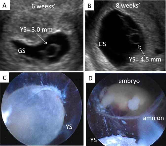
Figure 3.
(A) Ultrasound and hysteroscopic images of the yolk sac in a partial mole pregnancy (Karyotype: 69, XXY at microarray analysis). (A) Ultrasound picture showing an enlarged yolk sac at 6 weeks and 1 day of gestation; (B). Ultrasound picture showing an enlarged yolk sac at 8 weeks and 2 days of gestation; (C). Hysteroscopic view of the yolk sac at the time of pregnancy evacuation at 8 weeks and 2 days of gestation, after embryonal demise. (D) A portion of the yolk sac can be noted just outside of the amniotic sac, with the embryo within it, in the background. GS = gestational sac; YS = yolk sac.

