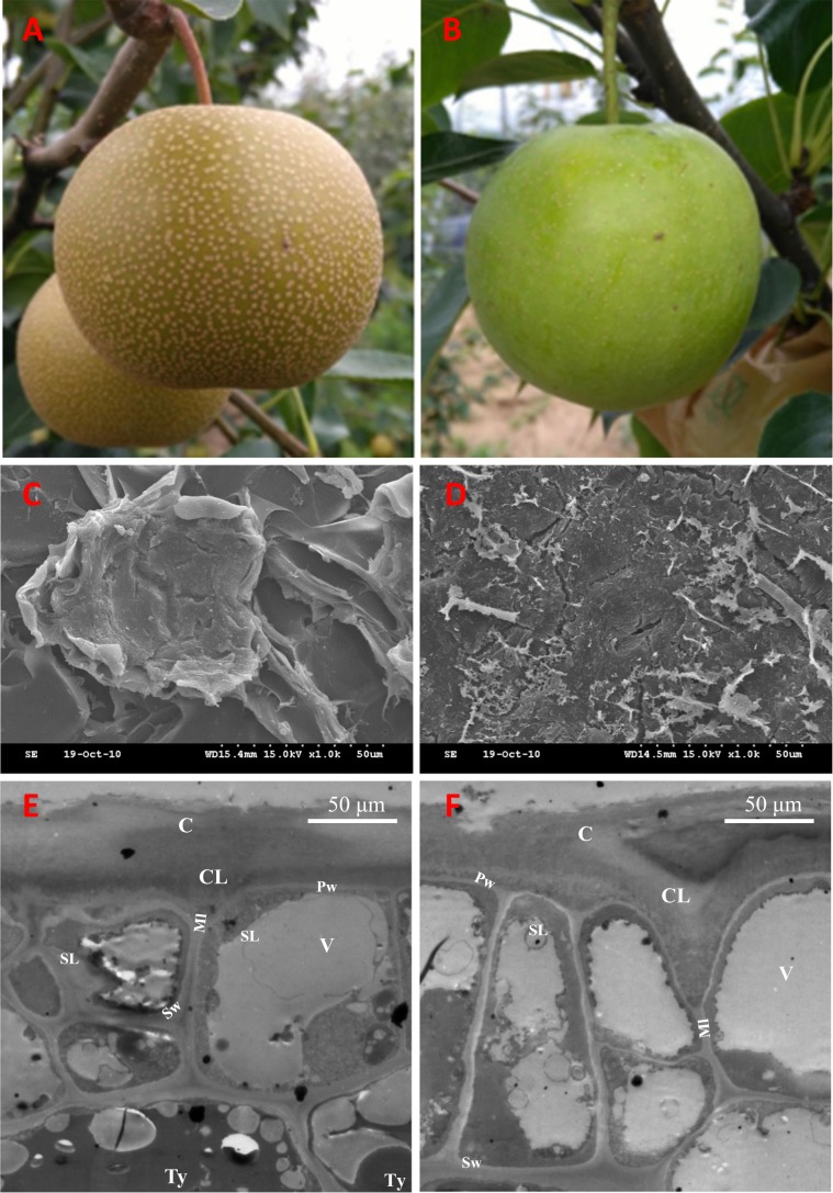
Fig. 1. Phenotypes of the russet and green fruit skin of sand pear.
The russet a and green b skin fruit of sand pear grown in an orchard. Scanning electron microscopy analysis of russet c and green d fruit surfaces showed that the skins of the two genotypes are covered by stacked suberized cells and cutin wax, respectively. Transmission electron microscopy analysis of russet e and green f fruit epidermal cells at the russet development stage showed that more tylosis (Ty) accumulated in the russet skin cells. C Cuticle; CL cuticular layer; Ml middle lamella; Pw primary wall; SL suberin layer; Sw secondary wall; V vacuole; Ty tylosis

