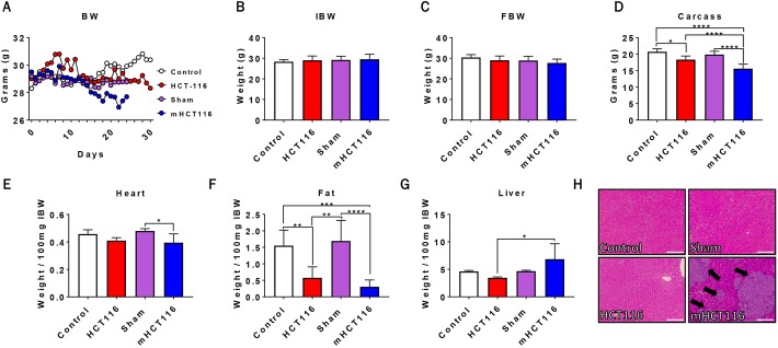
Fig. 1.
HCT116 subcutaneous and metastatic tumor hosts experience significant body weight and fat loss. (A-D) Body weight (BW) curves (A), initial body weight (IBW) (B), final body weight (FBW) (C) and carcass weights (D) of NSG male mice (8-week old) subcutaneously injected with HCT116 tumor cells (3.0×10⁶ cells/mouse in sterile PBS: HCT116) or equal volume of vehicle (control), or intrasplenically injected with HCT116 tumor cells (1.25×10⁵ cells/mouse in sterile PBS: mHCT116) or an equal volume of vehicle (sham) (n=5-8). (E-H) Heart (E), fat (F) and liver (G) weights normalized to IBW, and representative H&E staining of liver from control, HCT116, sham and mHCT116 mice (H). Black arrows indicate tumors and images were taken at 10× magnification. Scale bars: 100 µm. Data are mean±s.d. *P<0.05, **P<0.01, ***P<0.001, ****P<0.0001 (one-way ANOVA with Tukey's test).

