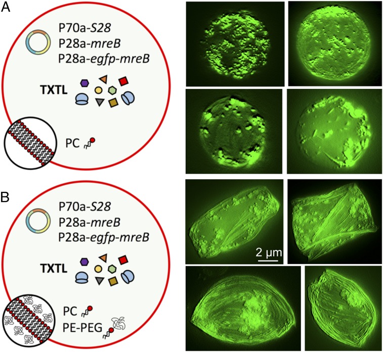
Fig. 1.
Image reconstruction from z scan in fluorescence microscopy. (A) Cell-free expression of mreB inside synthetic cells with a membrane composed of 100% PC. (B) Cell-free expression of mreB inside synthetic cells with a membrane composed of 99.33% PC and 0.66% PE-PEG5000. The cell-free reactions encapsulated inside the synthetic cells were programmed with the plasmids P70a-S28 (0.5 nM), P28a-mreB (2 nM), and P28a-egfp-mreB (0.2 nM). The fluorescence images of several liposomes (taken after 12 h of incubation) show MreB filament accumulating at the membrane in both cases. Deformations are observed when a lipid−PEG is used, as opposed to the case with no PEG at the membrane. These images were obtained from z-stack images reconstructed by the software MetaMorph. The scale bar is the same for all the images.

