Fig. 1.
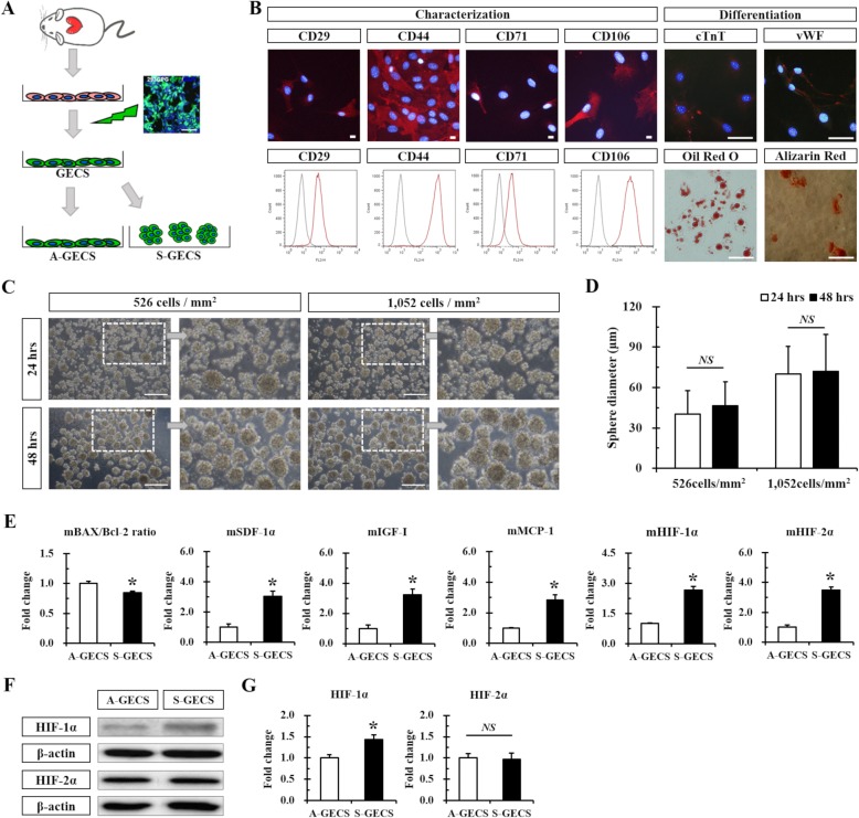
GECS generation and characterization. a Schematic diagram of GECS obtained from hTERT-immortalized Sca-1+ CSC lines. b Representative immunofluorescence images and flow cytometry of GECS. c Representative phase-contrast images of S-GECS produced on Poly-HEMA-coated plates 24 and 48 h after seeding. d Time-dependent increase in S-GECS diameter. n = 58 in each group. e Quantitative RT-PCR analysis of apoptotic, hypoxic, and growth factors in A-GECS and S-GECS for 48 h, each in triplicate. *p < 0.05 vs. A-GECS. f and g Western blot and quantification of HIF-1α and HIF-2α expression in A-GECS and S-GECS, each in triplicate. *p < 0.05 vs. A-GECS. All results are representative; scale bars represent 100 μm

