Fig. 2.
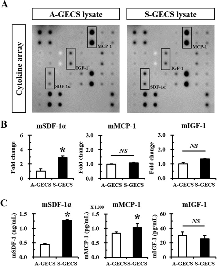
Cytokines/chemokine antibody array panels of S-GECS lysates. a Representative cytokine/chemokine antibody array panel of A-GECS and S-GECS lysates. b Comparison of highly expressed SDF-1α, MCP-1, and IGF-1 between A-GECS and S-GECS. Relative expression of cytokines and chemokines was measured by densitometry, each in triplicate. *p < 0.05 vs. A-GECS. (C) ELISA of SDF-1α, MCP-1, and IGF-1 between A-GECS and S-GECS, each in triplicate. *p < 0.05 vs. A-GECS

