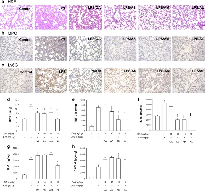
Fig. 7.
The effect of intravenous free OA and OA-loaded nanocarriers on LPS-induced lung injury in mice. a The lung histology (H&E staining) of LPS-challenged mice treated by free OA and OA-loaded nanocarriers. b The immunohistochemistry (MPO antibody staining) of LPS-challenged mice treated by free OA and OA-loaded nanocarriers. c The immunohistochemistry (Ly6G antibody staining) of LPS-challenged mice treated by free OA and OA-loaded nanocarriers. d MPO expression. e TNF-αexpression. f IL-1β expression. g IL-6 expression. h CXCL-2 expression. All data represent mean ± SEM (n = 6). *p < 0.05, **p < 0.01, ***p < 0.001 as compared to LPS-treated group without OA intervention

