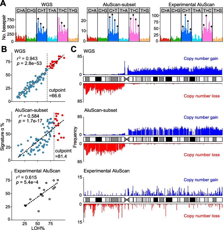
Fig. 7.
AluScan analysis of HCC patients. Comparison of (a) SNV profiles in WGS, AluScan-subset and Experimental AluScan sequences. (b) Correlations between Signature α%. (c) CN-gains and losses in WGS, AluScan-subset and experimental AluScan sequences on chromosome 8. The CNV frequencies in 350-kb windows were shown as bars above and below the chromosome ideogram for CN-gains (blue) and CN-losses (red), respectively.

