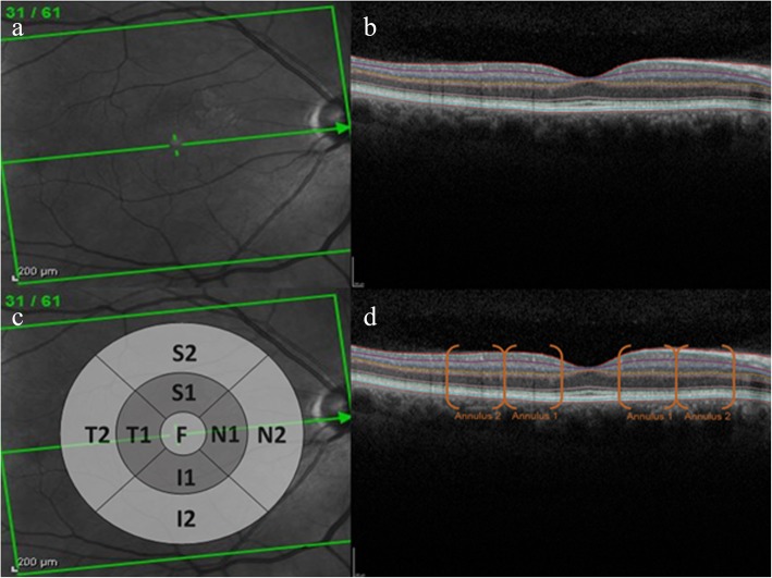
Fig. 1.
Retinal images and grid positions: a shows an image of the retina en face. b shows a cross-sectional image with differentiation of the retinal layers using the HAYEX software. The cross-section of the retina represents the layers directly behind the green line bisecting the en face image in panel A. The cross hairs in the left hand panel indicate the location of the foveal dip which can be seen as a depression in the centre of the image in panel B. c indicates the approximate position and size of the ETDRS grid used for reporting retinal thickness. Segment F is centred over the fovea. The annulus proximal to the fovea (annulus 1) comprises segments; S1 = superior 1; N1 = nasal 1; I1 = inferior 1; T1 = temporal 1. The annulus distal to the fovea comprises segments; S2 = superior 2; N2 = nasal 2; I2 = inferior 2; T2 = temporal 2. d highlights the locations where the ETDRS grid segments bisect the retinal image shown in panel B

