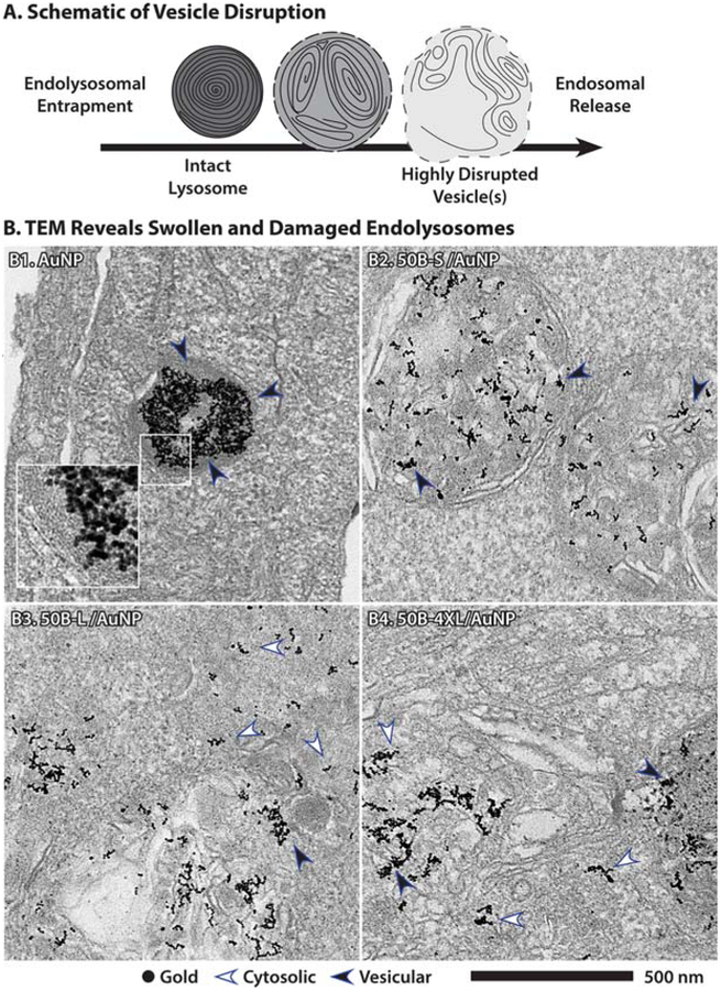
Figure 5.
Transmission electron microscopy confirmation of endosomal disruption as predicted by Gal8 recruitment. (A) Endolysosomes are multilamellar, electron-dense structures that become more swollen with weakly membrane active polymers, while highly membrane active polymers more fully disrupt endosomal membranes and induce a fragmented, multivesicular phenotype with incomplete or discontinuous membranes. (B) Transmission electron microscopy shows AuNPs traffic to electron-dense lysosomes, B1, where they are fully enclosed by a lipid bilayer (black arrow), while 50B-S induces a swollen vesicle phenotype, B2, although the membrane remains intact. Endosome disruptive polymers 50B-L and 50B-4XL induce widespread loss of endosomal membrane and release AuNPs into cytoplasm (white arrows), although some AuNP remains trapped in membranes (black arrows).

