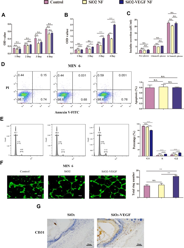
Figure 2.
The biomaterial properties are tested in vitro (pink as control, yellow as SiO2 NF, blue as SiO2-VEGF NF). (A) MIN-6 cells and (B) HUVEC cells are cultured with SiO2 NF or SiO2-VEGF NF for 1–4 days and cell viability are assessed by MTT assay. Absorbance was detected with a Microplate Reader at a wavelength of 570 nm; (C). The insulin release assay of MIN-6 cells is tested after they are pre-treated with SiO2 NF or SiO2-VEGF NF for 24 hrs. (D) Cell apoptosis is calculated by flow cytometry, and the percentages had no significant difference among these three groups. (E) HUVEC cell cycle assay indicated the cell percentage of S/G2 increased on SiO2-VEGF NF group for pre-cultured 24 hrs (SiO2-VEGF group, S=6.69; G2=11.0). (F).After suspended HUVECs were treated with SiO2-VEGF NF for 24 hrs, tube formation is observed by fluorescent microscope directly and the total ring number significantly increased. (G) CD31 staining (brown stain) verified the endothelia cell growth on SiO2-VEGF NF group (* represents P<0.05; ** represents P<0.01; *** represents P<0.001 and ns represents no significance, respectively).

