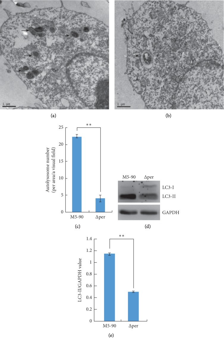
Figure 1.
Autophagy is inhibited in RAW264.7 cells infected with B. melitensis ∆per. (a, b) Transmission electron microscopy examination of RAW264.7 infected with B. melitensis M5-90 or B.melitensis ∆per for 4 h, respectively. Arrow indicates autolysosome. (c) The number of autolysosome of RAW264.7 infected with B. melitensis ∆per for 4 h was significantly decreased. (d) The levels of LC3-II/GAPDH. RAW264.7 cells were infected with B. melitensis M5-90 or B. melitensis ∆per for 4 h, respectively, and then the total cell lysates were prepared and subjected to immunoblot analysis using monoclonal anti-LC3-I antibody and polyclonal antibody against LC3-II. (e) The quantification of LC3-II/GAPDH levels in d with BandScan5.0 (n = 3). Scale bars are 1 μm. Data are mean ± SD from three independent experiments. ∗p < 0.05; ∗∗p < 0.01.

