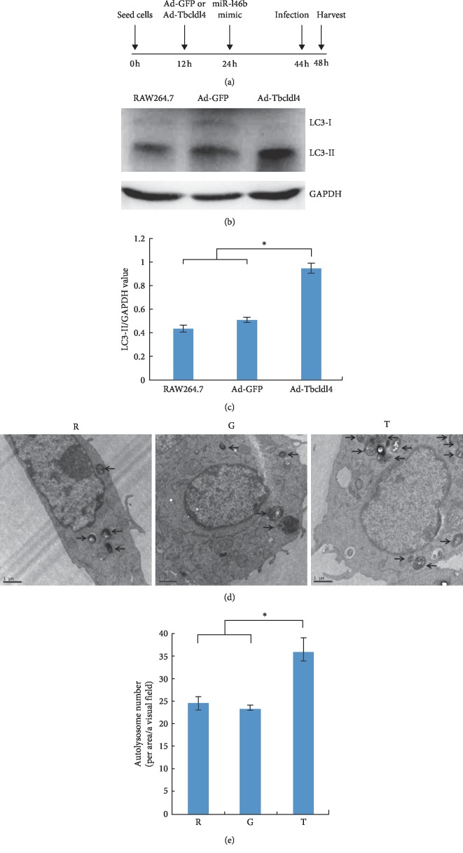
Figure 6.
Tbc1d14 contributes to miR-146b-5p-mediated inhibition of autophagy during B. melitensis infection. (a) RAW264.7 cells were seeded at 0 h, were infected with Ad-EGFP or Ad-Tbc1d14 after 12 h, were transfected with miR-146b mimic after 24 h, were infected with B. melitensis M5-90 after 44 h, and the total protein was extracted after 48 h. (b) Western blot analysis of the LC3-I and LC3-II protein expression. (c) The quantification of LC3-II/GAPDH levels in C with BandScan5.0 software (n = 3). (d) Transmission electron microscopy observation of the autolysosome in Ad-Tbc1d14 infected RAW264.7 group (Group T), Ad-EGFP infected RAW264.7 group (Group G) as negative control, and RAW264.7 group as blank control (Group R). (e) Statistical analysis for the number of autolysosomes in T, G, and R. Scale bars are 1 μm. Data are mean ± SD from three independent experiments. ∗p < 0.05; ∗∗p < 0.01.

