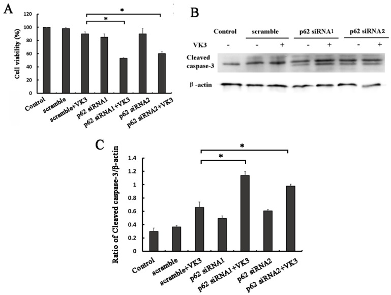
Figure 5.
p62 inhibition increased VK3-induced apoptosis in SKOV3/DDP cells. (A) SKOV3/DDP cells were transfected with p62 or control siRNA. After treatment with 15 µM VK3 for 16h, MTT assays was used to evaluate cell viability. Data are presented as mean ± SD, n = 3. *P < 0.05 compared with si-scrambled + VK3 treatment group. (B) SKOV3/DDP cells were treated as (A). And the expression of cleaved caspase-3 was analyzed by Western blotting. (C) The expression of cleaved caspase-3 in (B) was quantified. Data are presented as mean ± SD, n = 3. *P < 0.05 compared with si-scrambled + VK3 treatment group.

