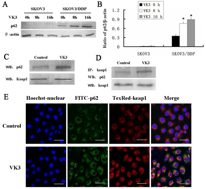
Figure 6.
The interaction between p62 and Keap1 increased with VK3 treatment in SKOV3/DDP cells. (A) Both cells were treated with 15 µM VK3 for 8 or 16 h. Cell lysates were subjected to immunoblot analysis with anti-p62. (B) The expression of p62 in (A) was quantified. Data are presented as mean ± SD, n = 3. *P < 0.01 compared with untreated cells. (C) SKOV3/DDP cells were treated as before, and total p62 and Keap1 were detected by western blotting. (D) Cell lysates were immunoprecipitated with anti-Keap1 antibody and immunoblotting was performed with anti-p62 and anti-Keap1 antibodies. (E) Cells were treated with 15 µM VK3 for 8 h. Immunofluorescence of p62 and Keap1 was detected by fluorescence microscopy (scale bar, 25 µm).

