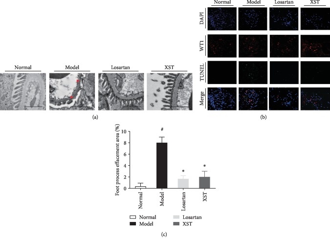
Figure 2.
Effects of XST on podocyte morphological changes and podocyte apoptosis in diabetic rats. Representative transmission electron photomicrographs (a) and semiquantitative ultrastructural analysis (c) in diabetic rats. Representative immunofluorescence labelling including the TUNEL assay, WT1, and DAPI (b) on frozen kidney sections. The cells with WT1 (red), TUNEL (green), and DAPI (blue) were identified as the positive apoptotic podocytes. Scale bar = 2 μm. Red arrowheads indicated podocyte foot process effacement. Results were expressed as the means ± SEM. #P < 0.05 vs. normal; ∗P < 0.05 vs. model.

