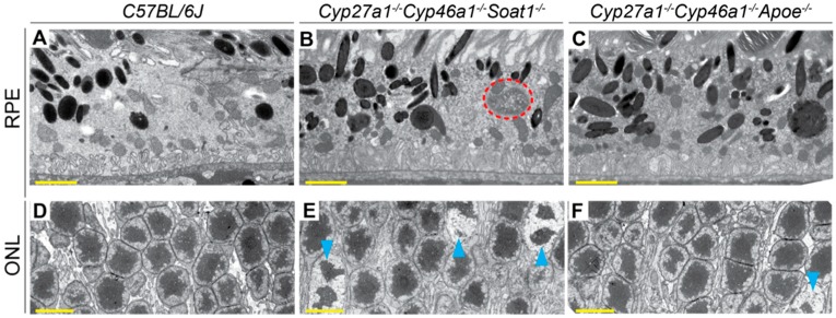
Figure 8.
Retinal ultrastructure as assessed by TEM. Cross sections through the retinal pigment epithelium (RPE) and outer nuclear layer (ONL) are shown. Orange dashed ellipse denotes undigested material; cyan arrowheads denote chromatin condensation. A, B, D, E: Images are representative of three male mice per genotype. C, F: Only one male animal was imaged. All mice were 7-months old. Scale bars: 2 μm (A,B,C); 5 μm (D,E,F).

