Figure 1.
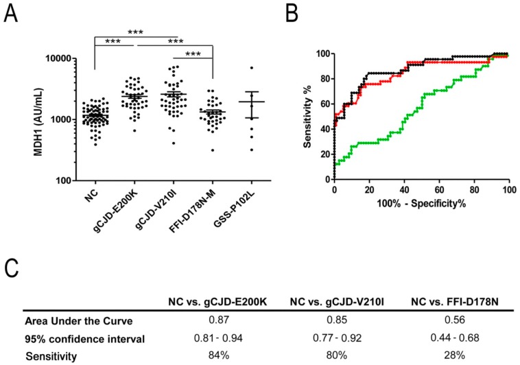
Detection of mitochondrial malate dehydrogenase 1 (MDH1) levels in cerebrospinal fluid (CSF) of genetic prion disease patients. (A) Patients with gCJD E200K (n = 45) and V210I (n = 46) contained a significantly increased level of MDH1 compared to fatal familial insomnia (FFI) cases (n = 36) and neurological controls (NC) (n = 71). GSS patients (n = 7) exhibited slightly higher MDH1 levels than FFI and controls, without statistical power due to a low number of samples. For comparison between groups we used Kruskal-Wallis test and Dunn’s post-hoc test. A p-value < 0.001 is considered as highly significant (***), <0.01 as very significant (**), <0.05 as significant (*), and ≥0.05 as not significant (ns). Values are indicated as arbitrary units (AU) per mL. Displayed are means ± SEM (standard error of the mean). (B) Receiver operating characteristic (ROC) curve analysis was performed to distinguish gCJD (E200K and V210I) patients and FFI from NC. Black, curve for gCJD-E200K; red, curve for gCJD-V210I; green, curve for FFI (C) area under the curve (AUC) values, corresponding to the area under ROC curves, and 95% confidence intervals are reported. Diagnostic sensitivity for gCJD patients was 0.85–0.87 AUC.

