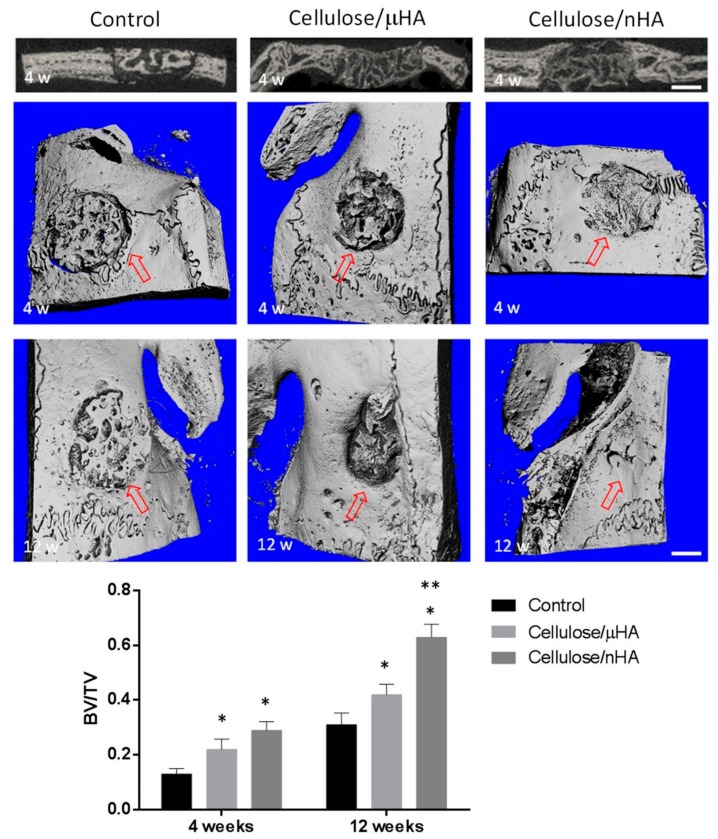
Figure 4.
Results of an in vivo rabbit calvarial defect model treated with cellulose-based composites, using HA-particles with either 20 µm (µHA) or 100 nm (nHA) in diameter, compared to untreated control. Figures above: 2D images and 3D reconstructions by means of µ-CT of the control and implanted composites after 4- and 12-week treatment, respectively. Margin of the initially created defect is highlighted by red arrows. Scale bar corresponds to 3 mm. Graph below: Ratio of bone volume (BV) to total volume of the created defect (TV). * indicate a significant deviation from control; ** indicate a significant deviation from cellulose/μHA. Reproduced from work in [127] with permission from John Wiley and Sons, 2019.

