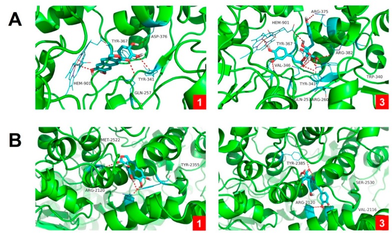
Figure 7.
Molecular docking results of compounds 1 and 3 with iNOS (A) and COX-2 (B) enzymes. Molecular docking simulations obtained at the lowest energy conformation, highlighting potential hydrogen contacts of 1 and 3, respectively (nitrogen is blue; oxygen is red; carbon is cyan; hydrogen is gray). For clarity, only interacting residues are labeled. Hydrogen bonding interactions are shown by dashes. These figures were created by PyMOL (Schrödinger, LLC, New York, NY, USA: version 1.3).

