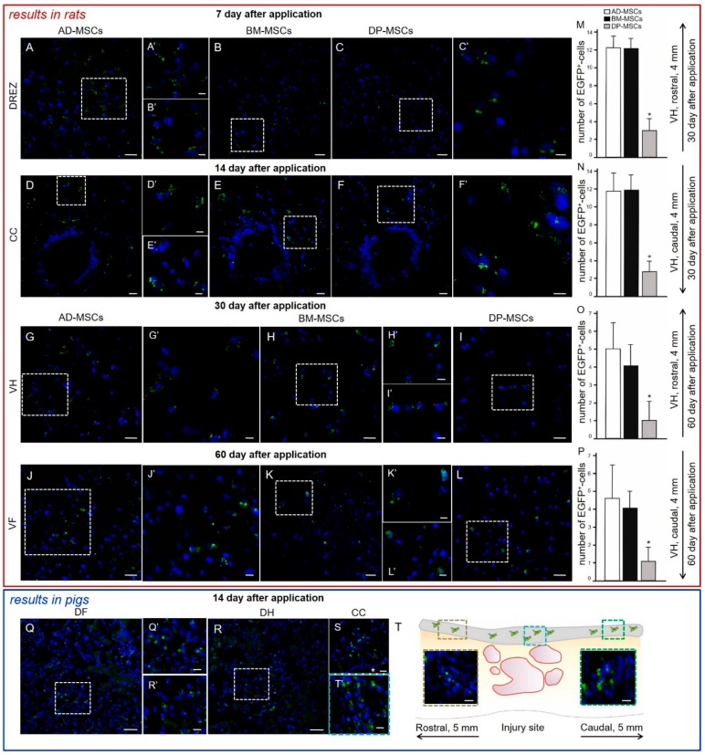
Figure 2.
Distribution and survival of MSCs encapsulated in FM after application into the area of SCI in rats (red box) and pigs (blue box). MSCs predominantly migrate through the posterior roots of the spinal cord. In the early periods (7–14 days post-transplantation) labeled by LV-EGFP (green) AD-MSCs, BM-MSCs, and DP-MSCs were located mainly in (A–C) DREZ and (D–F) central canal (CC) in rats. During late periods (30–60 days post-transplantation) labeled LV-EGFP cells were also observed in the (G–I) ventral horns (VH) and (J–L) ventral funiculi (VF) in rats. The (A–L) areas marked with a dashed boxed correspond to enlarged images A’–L’. Number of EGFP+-cells at days (M,N) 30 and (O,P) 60 at 4 mm rostrally and caudally from the injury site in VH of experimental groups with MSCs transplantation. At day 14, LV-EGFP MSCs (green) were mainly located in the area of DREZ, (Q) dorsal funiculi (DF), (R) dorsal horns (DH), and (S) CC in pigs. The Q and R areas marked with a dashed boxes correspond to enlarged images Q’ and R’, respectively. (T) MSCs distribution can be seen in the area of epidural fibrosis (gray line above spinal cord longitudinal section) according to the distance from injury site. MSCs were also detected in the area of epidural fibrosis, where more MSCs were found above the epicenter of injury and in the caudal direction, and less in the synechias above the rostral part of spinal cord. Asterisk indicates CC. Color dashed boxes (brown—rostral part; blue—epicenter of injury; green—caudal part) in scheme indicate corresponding area with MSCs distribution. Nuclei are stained with DAPI (blue). Scale bars: (A–L) 20, (H’) 10, and (A’–G’,I’–J’–L’) 5 μm; (Q,R) 50, (S) 20 and (Q’,R’,T’,E’, brown and green dashed boxes) 10 μm.

