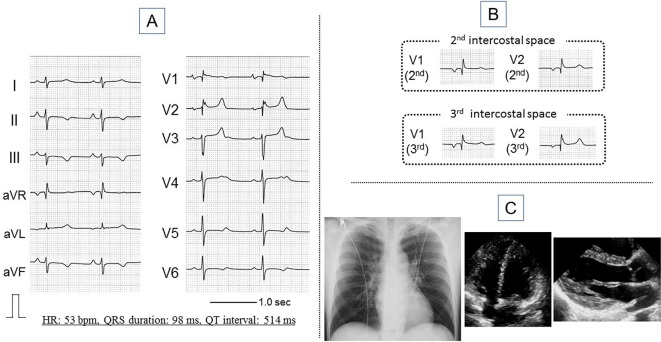
Figure 1.
Findings of the 12-lead electrocardiogram at rest (A, B), chest X-ray, and transthoracic echocardiogram (C). The electrocardiogram shows sinus rhythm, QT prolongation (QT interval: 514 ms, QTc interval: 485 ms), and mild saddleback-type ST elevation (A). Coved-type ST elevation is not observed, even in the second and third intercostal spaces (B). The chest X-ray and transthoracic echocardiogram findings are essentially normal (C). HR: heart rate

