Figure 2:
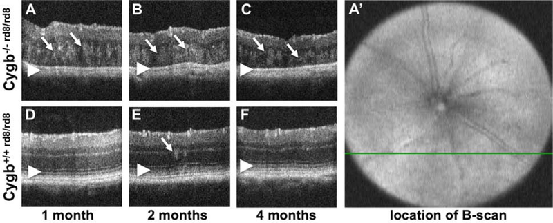
Optical Coherence Tomography of Cygb−/−rd8/rd8 mice shows outer retinal hyperreflective foci and loss of outer retinal bands. Areas of hyperreflectivity are seen as early as post-natal month 1 (A, arrows), and persist at postnatal month 2 (B, arrows) and 4 (C, arrows). Similar lesions are occasionally see in Cygb+/+rd8/rd8 animals (D,E,F) at these ages (arrow in E), but to a lesser degree. The outer retinal bands (D,E,F, arrowheads) representing the external limiting membrane, the inner segment/outer segment junction (aka ellipsoid zone), and the retinal pigmented epithelium are not discernable in Cygb−/−rd8/rd8 mice (A,B,C arrowheads). Also, the total thickness of the retina decreases with age in Cygb−/−rd8/rd8 mice (A,B,C). The b-scans shown here are taken from horizontal scans in the inferior retina. For example, the green line in A’ corresponds to the cross section in panel A.

