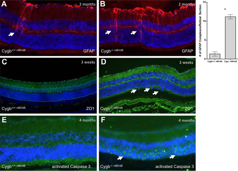
Figure 8:
Cygb deficiency results in disruption of the outer limiting membrane, reactive Muller glia, and cell death. Typical anti-GFAP staining is show in (A) Cygb+/+rd8/rd8 mice and (B) Cygb−/−rd8/rd8 mice at postnatal age 2 months. Quantification of GFAP-positive activated Muller glial complexes per retinal section is shown (*P<0.05 was considered significant, student’s t-test). The outer limiting membrane as stained by anti-ZO1 immunohistochemistry show relative preservation of this structure in (A) Cygb+/+rd8/rd8 mice, but with frequent areas of discontinuity seen in (B) Cygb−/−rd8/rd8 mice at postnatal age 3 weeks. Virtually no anti-activated Caspase 3 staining is seen at postnatal age 4 months in (E) the Cygb+/+rd8/rd8 retina, while robust staining is seen in the outer nuclear layer at this age in (F) Cygb−/−rd8/rd8 mice.

