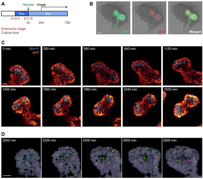
Fig. 4: Cell tracking of AE-rtTA;TRE-H2BGFP;Wnt11RFP embryonic kidney explants through high-resolution live imaging.
A: Pregnant AE-rtTA;TRE-H2BGFP;Wnt11RFP mice were given 2mg/ml doxycycline 24hr before harvesting at E11.5. Dissected kidneys were put on a Transwell filter plate with doxycycline-treated media (1mg/ml) and incubated at 37°C. After 24hr, kidneys were imaged every 5min for 48hr with z-stacking to capture the entire structure. B: E11.5 kidney post-dissection illustrating native GFP and RFP signal. C: Time-lapse imaging of kidney explants; see also Video S1. D: Automated tracking of cells from the time-lapse imaging. Line in B = 100μm; line in C, D = 30μm.

