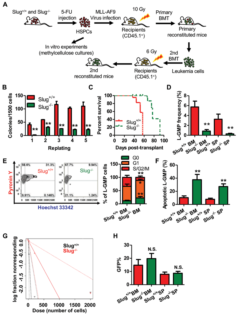
Figure 2. Slug deficiency impairs self-renewal of LSCs and delays MLL-AF9 leukemia onset.
(A) Experimental schema of MLL-AF9-driven AML. BMCs were isolated from Slug+/+ or Slug−/− mice treated with 150 mg 5-FU/kg BW for 6 days, transduced with MLL-AF9 retrovirus in vitro in order to generate AML cells, and transplanted into irradiated recipient mice (CD45.1). For primary transplantation, 1X10³ GFP+ cells were injected. For secondary transplantation, 5X10⁵ or 5X10⁴ GFP+ cells were injected.
(B) Colony-forming assay of Slug+/+ or Slug−/− AML cells (n = 3).
(C) Survival analysis of primary recipient mice. Median survival was 57 versus 77 days post-transplant for primary recipients of Slug+/+ or Slug−/− AML cells, respectively (P < 0.01, Mantel-Cox test; n = 5).
(D) Frequency of L-GMP in the BM and spleen (SP) from secondary recipients injected with 5X10⁴ Slug+/+ or Slug−/− AML cells at week 7 post-transplantation (n = 4).
(E) Cell cycle phase distribution of L-GMP cells in BM from secondary recipients injected with 5X10⁴ Slug+/+ or Slug−/− AML cells at week 7 post-transplantation (n = 4).
(F) Percentage of apoptotic L-GMP cells in the BM and SP from secondary recipients injected with 5X10⁴ Slug+/+ or Slug−/− AML cells at week 7 post-transplantation (n = 4).
(G) Limiting dilution assay of Slug+/+ and Slug−/− LSCs from secondary transplantation recipients. LSC/LICs frequencies calculated by ELDA software (n = 5).
(H) Quantification of homing AML cells at 16 h after transplantation of GFP+ gated AML cells from secondary recipients (n = 5).
Data are representative of two to three independent experiments. Excluding survival analysis, all data are represented as mean ± SD. Two-tailed Student’s t-tests were used to assess statistical significance (* P < 0.05; ** P < 0.01). See also Supplemental Figure 4, 5, and 7.

