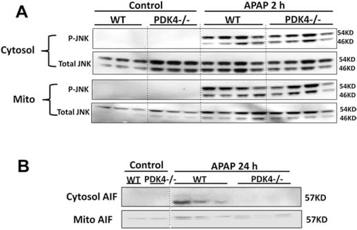
Figure 7:
JNK phosphorylation in liver cytosolic and mitochondrial fractions after APAP. Female wild type and PDK4−/− mice were treated with 600 mg/kg APAP or saline (control) and liver tissues were obtained from controls and 2 h post APAP. (A) Cytosolic and mitochondrial fractions were subjected to Western blotting for total JNK and phospho-JNK. (B) Mitochondrial release of apoptosis-inducing factor (AIF) at 24 h after APAP. Samples from 3–4 animals per treatment group were analyzed.

