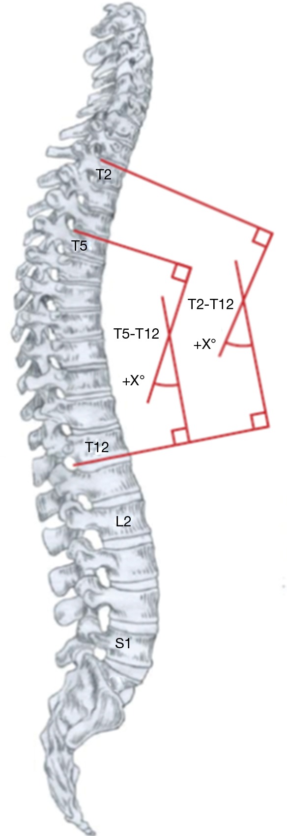
Figure 3.

Thoracic sagittal alignment. Thoracic kyphosis is measured from the upper-endplate of T2 to the lower endplate of T12 using the Cobb method. The upper thoracic spine is the most difficult to image, and it is a common occurrence not to have a clear shot of T2 on a radiograph, due to superposition of overlapping structures. Proximal thoracic kyphosis is measured from the upper-endplate of T2 to the lower endplate of T5. Mid/lower thoracic kyphosis is measured from the upper-endplate of T5 to the lower endplate of T12. Image courtesy of Scoliosis Research Society, from (28).
